EEG & fMRI
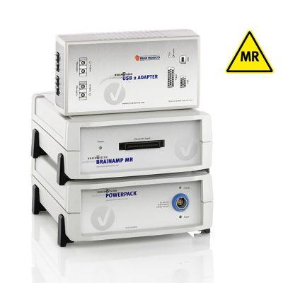
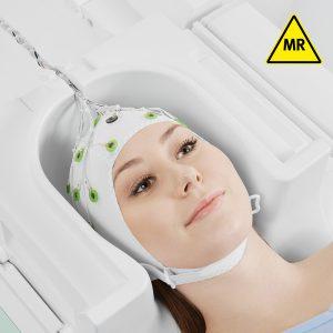
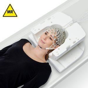
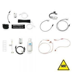
EEG and fMRI measure electrical and hemodynamic changes in the brain, respectively and are complementary in terms of spatial and temporal accuracy. Simultaneous acquisition of EEG and fMRI requires extra considerations for both data signal quality and participant safety. Our MR conditional amplifiers are officially certified by all major scanner vendors. These amplifiers can be combined with additional physiological sensors like accelerometers and GSR. Record your data fully synchronized to the scanner clock and, to ensure data quality during a recording, perform real-time processing of your data with our online viewing software. Our analysis software makes use of specialized transformations for offline pre-processing of scanner-related artifacts, as well as advanced analysis.
Recommended solutions
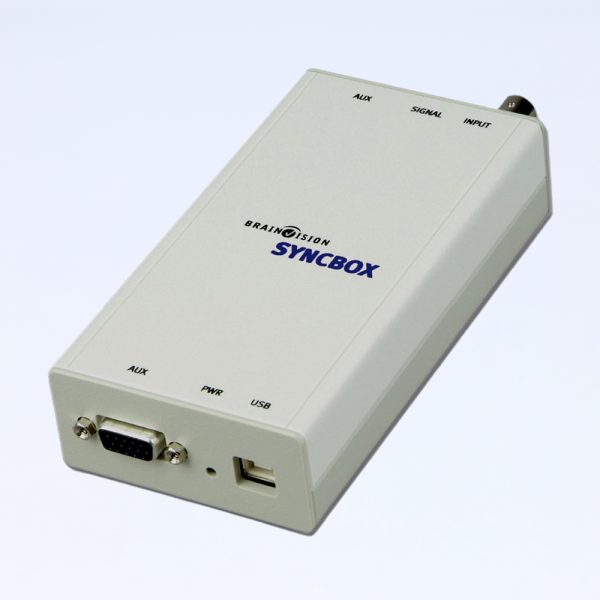
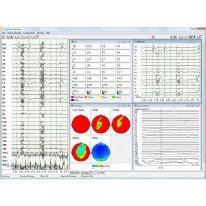
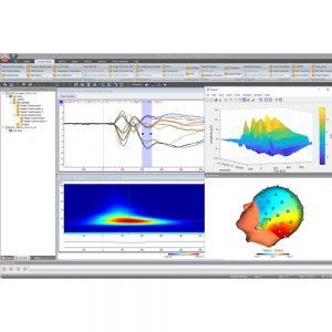
Below you will find some of our recommended solutions for this application field. Depending on your research paradigm, other solutions may be more appropriate. For the optimal setup for your research goals, please contact your local distributor.
More resources
Reference labs
We enjoy the close contact we have with research groups using Brain Products equipment. In fact, these collaborations often help us improve our solutions, not just for one specific application field, but for all. We greatly appreciate the ongoing collaboration with these labs.
Publications
Here are some papers related to this application field (published by the above mentioned reference labs):
- Rajkumar, R., Régio Brambilla, C., Veselinović, T., Bierbrier, J., Wyss, C., Ramkiran, S., Orth, L., Lang, M., Rota Kops, E., Mauler, J., Scheins, J., Neumaier, B., Ermert, J., Herzog, H., Langen, K.-J., Binkofski, F. C., Lerche, C., Shah, N. J., & Neuner, I. (2021). Excitatory–inhibitory balance within EEG microstates and resting-state fMRI networks: assessed via simultaneous trimodal PET–MR–EEG imaging. Translational Psychiatry, 11(1), 1–15. https://doi.org/10.1038/s41398-020-01160-2
- Bagshaw, A. P., Hale, J. R., Campos, B. M., Rollings, D. T., Wilson, R. S., Alvim, M. K. M., Coan, A. C., & Cendes, F. (2017). Sleep onset uncovers thalamic abnormalities in patients with idiopathic generalised epilepsy. NeuroImage: Clinical, 16, 52–57. https://doi.org/10.1016/j.nicl.2017.07.008
- Mayhew, S. D., & Bagshaw, A. P. (2017). Dynamic spatiotemporal variability of alpha-BOLD relationships during the resting-state and task-evoked responses. NeuroImage, 155, 120–137. https://doi.org/10.1016/j.neuroimage.2017.04.051
- Shah, N. J., Arrubla, J., Rajkumar, R., Farrher, E., Mauler, J., Kops, E. R., Tellmann, L., Scheins, J., Boers, F., Dammers, J., Sripad, P., Lerche, C., Langen, K. J., Herzog, H., & Neuner, I. (2017). Multimodal Fingerprints of Resting State Networks as assessed by Simultaneous Trimodal MR-PET-EEG Imaging. Scientific Reports, 7(1). https://doi.org/10.1038/s41598-017-05484-w

 Multimodal Integration Group (MIG)
 Institute of Neuroscience and Medicine – 4 (INM-4)